The best health care doesn't come from equipment or buildings; it comes from people. At the Breast Health Center, we combine advanced technology and a full array of treatment options with the most crucial element – specialized expertise and compassion. Every day it is our privilege to evaluate and treat women with the care and dignity they deserve. This is our mission and why we are the best choice for breast health services.
The Breast Health Center at Kent is proud to be a Women & Infants Hospital and Warren Alpert Medical School of Brown University Affiliate.
The Kent Breast Health Center delivers the highest quality services ranging from breast cancer screening, cancer treatment, managing benign breast issues, and helping with lactation problems. It is staffed by a collaborative and multidisciplinary team made up of specialty-trained breast health physicians, experienced nurses, and patient navigators.
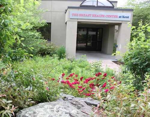
The Breast Health Center at Kent Hospital
455 Toll Gate Road
Warwick, RI 02886
![]()
To schedule a doctor’s appointment
P: (401) 736-3737
For mammograms
P: (401) 336-6699
Breast Health Center
Women & Infants Hospital
668 Eddy Street
Providence, RI 02903
P: (401) 274-1122, ext. 47300
P: (401) 453-7540
F: (401) 921-6925
Care New England Medical Group Primary Care and Specialty Services
Pawtucket Memorial Campus
111 Brewster Street
Pawtucket, RI 02860

Care New England Center for Health
49 South County Commons
South Kingstown, RI 02879
The key to surviving breast cancer is early detection and the only way to ensure early detection is to see a doctor regularly for mammography screening.

The Kent Breast Health Center strives to ensure you have seamless and coordinated access to an array of diagnostic, consultative, therapeutic, and supportive services, including:
Breast Imaging & Biopsies
Specialty Clinics
Multidisciplinary Breast Tumor Board
Experts review all breast cancer treatment plans from Kent Hospital and Women & Infants Hospital Breast Health Centers for the best and most current recommendations.
Breast conserving surgery removes the cancer, but not the entire breast. There are many types of incisions that can be used and each patient’s surgery will be uniquely planned. This type of surgery has many names including but not limited to lumpectomy, partial mastectomy, segmental resection, and quadrantectomy, but they all mean essentially the same thing—the surgical removal of the tumor and a small amount of normal breast tissue around it.
Surgeons at The Breast Health Center are trained in the most advanced oncoplastic surgery techniques for removing the cancerous tissue while also reforming the breast to avoid any deformity and preserve or improve a normal shape when possible. They also pair with plastic surgeons as necessary to ensure the best cosmetic results possible.
Kent Hospital is pleased to offer a breast cancer support group for all patients with a current or past history of breast cancer. It meets every 3rd Wednesday of the month from 2:00 p.m. - 3:00 p.m. It is an informal, ongoing group, bringing people together to share their experiences and problems related to cancer and to provide one another with comfort and help. Please use the Breast Health Center entrance located next to Kent's Emergency Department, 455 Toll Gate Road, Warwick, RI.
The Breast Health Center at Kent Hospital
Conference Room
Women & Infants Hospital
Breast Health Center
668 Eddy St.
2nd Floor
Providence, RI 02903
P: (401) 430-7111
Medical Director, The Breast Health Center at Kent Hospital
Jamie B. Patterson is the Medical Director of Kent Breast Health Center. She is a fellowship-trained breast surgeon that advocates for the highest quality and most comprehensive breast care for women. She earned her medical degree at the University of Nevada School of Medicine as a junior recipient of the Alpha Omega Alpha award, followed by training in obstetrics and gynecology at the University of California Irvine Medical Center with numerous awards for surgical excellence. She continued on as Assistant Clinical Professor and academic teaching faculty for the program before completing fellowship training in breast surgical oncology at the University of Southern California/Hoag with renowned breast surgeon, Dr. Melvin Silverstein, who pioneered oncoplastic breast surgery in the United States.
Director, Medical Oncology
Chief, Cancer Program at Kent Hospital
Dr. Sakr joined the Program in Women's Oncology in 2007. Dr. Sakr received a degree in medicine from The Lebanese University in Lebanon and completed his fellowship in hematology and oncology at the University of Pittsburgh Medical Center in Pennsylvania.
Director of Plastic & Reconstructive Surgery
Dr. Erik Hoy earned his medical degree from Rutgers-New Jersey Medical School. He is a board-certified plastic surgeon specializing in aesthetic surgery of the breast and body, reconstructive breast surgery, autologous fat grafting, and anatomic breast implants, as well as soft-tissue reconstruction after cancer resection.
Breast Surgeon
Micaela Weaver is a fellowship-trained breast surgeon and general surgeon. She received her Doctor of Osteopathic Medicine degree at West Virginia School of Osteopathic Medicine. Dr. Weaver completed her general surgery residency at Flushing Hospital Medical Center in Queens, New York, and her breast surgical oncology fellowship training at Women & Infants.
Medical Oncologist
Ambreen Ijaz, MD, graduated from Fatima Jinnah Medical College, Punjab University Pakistan, and pursued her internal medicine residency at Brown University. Due to her profound interest in hematology/medical oncology, she joined Tufts University for her fellowship which she completed in 2011.
Plastic & Reconstructive Surgeon
Affiliated with Brigham and Women’s Hospital
Dr. Christian offers a broad range of reconstructive and cosmetic plastic surgery and hand surgery services to adults and children of all ages. He is board-certified in plastic and reconstructive surgery and has additional training and certification in surgery of the hand.
Director of Breast Imaging
Dr. Khalil graduated from the American University of Beirut. She completed her residency at Bridgeport Hospital, Connecticut and completed a fellowship in breast and body imaging at Memorial Sloan Kettering Cancer Center. Dr. Khalil is the Director of Breast Imaging for Kent Hospital. She is also an Assistant Professor of Diagnostic Imaging at The Warren Alpert Medical School of Brown University. Dr. Khalil’s special interests are breast and Ob/Gyn imaging.
Genetic Counselor
Nurse Manager & Cancer Navigator
Nurse Navigator
Nurse Practitioner
A risk factor is something that indicates a woman is more likely than average to develop breast cancer. They are not harmful in themselves. In fact, a woman's risk of developing breast cancer remains low even if she has several risk factors. About 85 percent of women who develop breast cancer do not have significant risk factors.
A history of breast cancer in immediate female relatives, especially at a young age, may indicate a genetic predisposition and constitutes a major risk factor in five to 10 percent of breast cancer patients.
The two most significant risk factors are being a woman and getting older.
The third most significant risk factor is having a personal history of breast cancer. A diagnosis of atypical hyperplasia after a biopsy may increase the risk of developing invasive breast cancer. Women who have been diagnosed with other cancers such as colon, ovarian, or endometrial may be at an increased risk.
Fibrocystic changes do not increase a woman's risk of developing breast cancer since almost all women have some aspect of this condition. Only breast duct lining changes showing abnormalities increase risk. This diagnosis can only be made through a biopsy.
Not all families' histories of breast cancer increase the risk to unaffected family members. The risk increases when a first-degree relative (sister or mother) has developed breast cancer at an early age, particularly in their 30s before menopause, or where multiple family numbers have breast cancer. Both maternal and paternal sides of the family should be considered when looking at family history. The risk seems to increase when cancer has appeared in both breasts of relatives.
Eighty-five to 90 percent of all breast cancers are diagnosed in women with no family history. Breast cancer is individual and based on the interaction between a woman's environment and heredity. American diets, which are high in animal products, contribute to an increase in breast cancer in contrast to vegetarian diets. No evidence exists, despite many scientific studies, of increased risk from exposure to herbicides, pesticides, hair dye, deodorants, or other chemicals in our American lifestyle.
Radiation treatments in the vicinity of the breasts for the treatment of childhood cancers increase risk greatly.
Sometimes women with a higher risk of developing breast cancer may benefit from more thorough screening. Examples of higher risk would include women who carry a strong genetic predisposition to breast cancer, such as carriers of a BRCA mutation, women with a history of radiation to the chest, and those who have three or more first-degree relatives with breast cancer. Breast MRI may be offered to such women in addition to annual mammography to help find cancer at the earliest, most curable stages. Breast MRI requires the use of intravenous dye to look at both the anatomical structure and blood flow characteristics of the breast. Because its findings are less specific, breast MRI may find other lesions that require additional tests but ultimately prove benign. Therefore, it is not a screening device for “average risk” women in the general population.
Several other roles for breast MRI are evolving and further demonstrate its importance as an imaging device. Breast MRI can be helpful in defining the extent of disease or second breast cancer at the time of initial diagnosis if recommended by your doctor. It can influence decisions regarding the most appropriate surgical procedure especially in women with “dense breasts,” which can make mammography more difficult. In the setting of locally advanced breast cancer, when chemotherapy or hormonal therapy is given before definitive surgery, breast MRI can also be critical in evaluating response to treatment before surgery.
Patients should discuss with their providers if Breast MRI is right for them. To schedule an appointment with a provider in our HOPE high-risk clinic, please call (401) 736-42600.
Women detect most lumps in their breasts themselves by performing breast self-exams (BSE). Performing regular self-exams will familiarize you with how your breasts feel normally so you will be able to more easily recognize changes.
Even though most breast lumps are not cancerous, finding one often causes anxiety. If performing a BSE causes you anxiety, speak to your physician who might suggest you stop them and have exams in the office only. Do not feel guilty. There is no evidence that self-exams decrease breast cancer deaths.
The best time to do a monthly BSE is right after your period when the breasts are no longer tender or swollen. Plan to perform the exam roughly the same time each month.
This will significantly impact your prognosis (your long-term chance of survival). Before surgery, your stage is an estimate also known as a ‘clinical’ stage because your final stage is often not definitively known until after your cancer is removed. At that time, your doctor will assign you a ‘pathologic’ stage once all the exact details of your cancer are known such as the true size and lymph node status.
Fear is a powerful motivator, but not as strong as you are! Many women initially react to a diagnosis of breast cancer with the reaction that they want both their entire breasts removed. But many women can safely save their breasts. It’s important to have a discussion with your doctors about whether removing your breast/s will change your prognosis—many times it won’t. Newer research tells us if a woman is able to save her breast safely, she will be less likely to experience depression, anxiety, and sexual dysfunction after her treatment. It’s the responsibility of your doctor to address all of those things, and all of the health implications that are important to you as a person, not just your cancer. Newer surgical techniques, like Oncoplastic Surgery (cancer surgery combined with plastic surgery techniques to improve immediate cosmetic outcomes), can be used in most cases to help preserve the breast shape and contour even with larger tumors if needed. Medications can also be given prior to surgery by a medical oncologist, called neoadjuvant therapy, in order to attempt to shrink your tumor and help minimize the amount of surgery you’ll need. You should feel empowered to ask if either of these options could be good choices for you. Certainly, the ultimate decision is intimately personal and a choice best navigated between you and your doctor. Some women feel so nervous after a breast cancer diagnosis that the thought of ever having to undergo another mammogram after treatment may be enough for them to want to undergo a mastectomy—that’s a valid reason, a personal choice. But medically-speaking, a woman should almost always be presented with at least an attempt to save her breast.
Once upon a time, all women with breast cancer were treated with chemotherapy. Now-a-days that’s not always the case. Breast cancers are unique, they’re made from your own DNA even if they’ve gone haywire, so they’re all different and you can’t really compare someone else’s treatment with yours. As doctors, we categorize breast cancers into groups based on receptors, or signaling proteins on the surface of cancer cells that respond to certain hormones or medications, they give us a better idea of how each will respond to different treatments. Certain types of breast cancer, like those with Her2 receptors or no receptors (often called “triple-negative” cancers), are still best treated with chemotherapy (though the exact medicines are even more specific), but the majority of breast cancers can respond to hormonal therapy. Hormonal therapy, or endocrine therapy, is typically a pill you take once a day that stops your female hormones (estrogen and progesterone) from stimulating your cancer cells to grow. All women, and even men, have some female hormones, even after menopause; they’re just made in lower concentrations in your adrenal glands and fat cells instead of your ovaries. By blocking or destroying those hormones, the medicine is essentially starving your cancer cells. If you have a lower-risk type of cancer, your doctor may recommend that endocrine therapy is all you need.
Most breast cancers are not inherited through your family, only about 5-7% of them are. That can be surprising to many people. The majority of the hereditary cases result from mutations, or changes, in the BRCA genes, which cause a condition known as Hereditary Breast and Ovarian Cancer Susceptibility Syndrome (HBOCS). Other genes, some known but many of which are still unknown, may also cause hereditary or familial cancers. Gene mutations result from damage to the cell’s DNA before you were born and therefore are inherited from a parent. This is why most individuals with breast cancer gene mutations will have a family history of breast cancer in young women (<50 years old), multiple affected family members, and/or ovarian cancer. Mutations within the BRCA genes are especially common in the Ashkenazi Eastern European Jewish population. Many treatment teams offer blood tests to identify gene mutations if indicated even before surgery. These tests will help you determine if your breast cancer is hereditary (passed in your family) or sporadic (unrelated to your family). It can also affect your chance of developing second breast cancer later in life and of course your family members’ chances of developing breast cancer.
A new diagnosis of breast cancer is shocking and stressful. You may feel any number of vastly different, but all valid, emotions, and most likely no one, not even your partner, best friend, or other survivors, will quite understand what you’re going through. How could they? But just because you may feel alone (or not!) doesn’t mean you can’t let people help you. Support groups or counseling are always good places to talk to someone, but even if you don’t want to talk about it, there’s always something you can let someone who cares about you do. Maybe you just want a home-cooked meal. Or someone to clean your house for you, watch the kids for a few hours, walk with you, play music, or even go fishing with you. (It doesn’t even have to be someone you know!
See CleaningforaReason.org, NannyAngelNetwork.com, MusicBoxAngels.org, or CastingforRecovery.org )
Sometimes it just helps to take your mind off things. And don’t forget, you deserve a break.
For questions, call (401) Care Now or visit us here to make an appointment. We're here to help.
Don’t panic. You don’t know what you’re dealing with until you’ve seen your doctor and have more information. Women often find lumpy parts of their breasts that can be attributed to normal hormonal changes, dense breast tissue, cysts, benign growths, or even parts of their normal anatomy like their ribs underneath their breast tissue or the edge of their breast, called the inframammary fold.
These normal changes can often be mistaken for a lump and can cause some real anxiety—so make sure you see your doctor right away first so they can help you determine if what you feel is really a lump.
Your physician may also want you to undergo breast imaging with a mammogram and/or ultrasound to help make that determination. These are important tests that will give you and your doctor a better idea of what you’re feeling.
Other associated symptoms will also help your doctor: whether the lump has changed in size or shape if it’s associated with a rash or other changes to your skin, or whether you’ve had have any pain or nipple discharge on that side.
Other risk factors such as your age and family history will also be taken into account. If the lump can be explained, you may not need anything further than follow-up breast exams with your doctor, or repeat imaging to ensure nothing changes. But if your doctor is worried or imaging shows a concern, you’ll most likely be asked to undergo a needle biopsy for a more definitive diagnosis.
If you’re ever concerned, you can always request your doctor consider one of these imaging or biopsy tests to be sure.
The main thing to remember is, not all lumps are cancer.
In fact, many are non-threatening or even normal, so while you definitely want to get evaluated, be sure you speak with your doctor before you let your worry keep you up at night.
Don't let the fear of the unknown stop you from taking the first steps. Our comprehensive care teams at both Kent Hospital and Women & Infants are here to help you in your journey, wherever that may take you. To schedule an appointment for routine screening, call (401) Care Now to find a location nearest you.
While 1 in 12 women will develop breast cancer in their lifetime, the majority of cases are not believed to be due to inherited genetic factors. However, approximately 10-15% of breast cancers are due to inherited genetic changes known as mutations. BRCA1 and BRCA2 are the two genes most commonly associated with hereditary breast cancer. Inherited mutations in these genes increase the risk for breast (55-85% lifetime risk), ovarian (23-55% lifetime risk), pancreatic (up to 5% lifetime risk), and prostate (15-25% lifetime risk) cancers.
More recently multiple additional genes have been identified that can also result in hereditary breast cancer and are available for testing. Genetic testing, which is performed on a small sample of blood or saliva, can identify individuals who carry these genetic changes in order to best estimate cancer risk for both themselves and their family members.
If you or a close family member have any of the following risk factors you may benefit from genetic testing:
Understanding whether you carry a genetic mutation can allow you, along with your doctors to create a personalized screening plan that can help to reduce the chance of developing cancer or identify it earlier. The cost of genetic testing has decreased substantially in recent years, and the vast majority of people with risk factors will have little to no cost for testing.
A meeting with a cancer genetic counselor can help you to understand if genetic testing is right for you. Together with the genetic counselor you can review your personal and family history and discuss the option of genetic testing if appropriate. Once the results are available, the genetic counselor will help to explain the implications for both you and your family, as well as provide a thorough cancer risk assessment and medical management plan to your treating physician team.
Visits with a cancer genetic counselor are available at The Breast Health Center at Kent Hospital and can be scheduled by calling (401) Care Now.
Mammography looks for a “dense cancer” in the background of the breast tissue. Breast tissue is made of fatty and glandular tissue. The ratio of glandular tissue to fatty tissue is the breast’s “density” and is somewhat related to one’s size, shape, and menopausal status, and can be hereditary. Mammographic density is not associated with how “lumpy” or dense the breast feels.
Naturally, the denser the tissue is, the harder it will be to find cancer that is identified due to its own “density.”
“I often say it is like trying to see the sun in a cloudy sky, with the sun representing cancer, and the clouds the density of a breast,” says Jennifer Gass, MD, of The Breast Health Centers at Women & Infants Hospital and Kent Hospital.
Breast density is a twofold issue for women. It makes it harder to read a mammogram and it has been associated with a modest increase in one’s risk for breast cancer. Why mammographic density causes an increased risk for breast cancer is an area of active research.
In the last several years, many states have enacted legislation that mandates women be informed of their breast density so they can make decisions about additional breast imaging. Three options are available at most institutions to examine dense breasts:
3D tomosynthesis, which is a layered breast imaging tool associated with a reduced call-back rate for false positives and earlier detection of cancer.
Breast ultrasound, a radiation-free tool that evaluates the breast and can find cancers a mammogram may have missed.
Breast MRI, a radiation-free test that is paired with contrast injection and takes 30 to 45 minutes. It is the most sensitive test, and the most expensive.
“Breast MRI has not been validated as an appropriate screening tool for women of average risk, or with risk based on breast density alone,” Dr. Gass explains.
Susan Koelliker, MD, director of diagnostic imaging at Women & Infants, adds that “Studies have shown that there is variability between different radiologists, and differences even when read by the same radiologist. The question is should this assessment become more standardized and potentially computer-assisted.”
However, knowing that breast density is often not uniform in the breast, she adds a computer-generated method might misrepresent the nuance. A combination of human and computerized efforts might seem an ideal synergy but such systems are expensive and would need to be reimbursed.
“Before we recommend embarking on yet another layer of costly technology for patients, we need to be sure there are robust outcomes that warrant this expense,” Dr. Gass says.
If you think you might be at risk of developing breast cancer, we encourage you to speak with your doctor about undergoing a risk assessment by making an appointment at The Breast Health Center at Kent Hospital or at Women & Infants, please call (401) Care Now or visit us here to schedule your screening today.
Copyright © 2023 Care New England Health System
 Same Day Mammograms >>